
True-To-Life Cardiac Anatomy and Echo Simulation
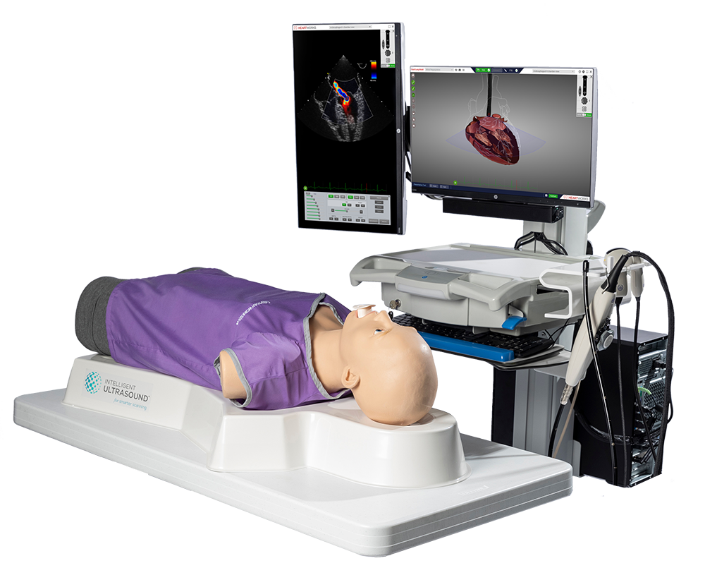
HeartWorks delivers comprehensive education in cardiac anatomy and echocardiography. Developed by leading clinicians in cardiac anesthesiology, it features a fully interactive 3D heart with 135 detailed intracardiac structures, over 30 pathology cases, and an integrated anatomy textbook, offering an unparalleled learning experience. Users can explore cardiac structures in detail, rotating and slicing the heart in any direction to deepen their understanding of ultrasound imaging.
Tailor your training with HeartWorks’ flexible learning pathway, covering Transthoracic (TTE), Transesophageal (TEE), and 3D echocardiography. Expand your curriculum with eLearning and on-system testing, or integrate HeartWorks with BodyWorks for a complete POCUS and echocardiography solution.
HeartWorks
Features and Benefits
- 30 patient cases, including normal heart and lungs, various diseased states, and key cardiac devices. All of which can be interrogated with Color, PW & CW Doppler, M-Mode.
- Best practice imaging views for 20 TTE and 28 TEE imagining planes, meaning trainees can learn to acquire these views without the need for tutor supervision.
- Teach your way with a cardiac learning pathway tailored to suit your curriculum. Build impressive presentations on cardiac function or anatomy, create tailored on-system assessments, or take learning on-the-go with supportive eLearning courses.
- Comprehensive Transthoracic Echocardiography (TTE) scanning with accurate, palpable anatomical landmarks to aid probe positioning and image acquisition.
- True-to-life Transesophageal Echocardiography (TEE) examinations with realistic controls for ante and retroflexion, lateral flexion and, omniplane rotation.
- Get an unparalleled understanding of complex cardiac anatomy and pathology with the 3D echocadiogreaphy add-on, teaching the principles of real-time 3D image acquisition and manipulation.


Download the HeartWorks Brochure
Learn more about the HeartWorks simulator.
Contact Us
For full specifications and pricing or to request a demo, please fill out the form below.